The doctor said he needed expensive software, the patient made a tool to see the results of his own 3D jaw x-ray with Claude, this is the prompt

Executive Summary:
- Independent Solution: Paolo Rosson managed to open and visualize 800 RAW Export Dicom files from his 3D jaw x-ray without buying expensive clinical radiology software.
- Claude Code Execution: Utilizing the capability of the AI Coding Agent Claude Code from Anthropic, it composes a self-contained medical image-based image displayer application that works completely offline.
- Medical equivalent features: This AI-made viewer presents a synchronization of 3 points of view (axial, coronal, sagittal) as well as precise 3D reconstruction rendering in millimeter units.
The accessibility of medical record data is often bumped by a complex proprietary file format or industry standard. This experience was experienced directly by a patient named Paolo Rosson. After undergoing a 3D dental and jaw X-ray scan procedure, the clinic handed over a folder containing 800 DICOM standard raw files (Digital Imaging and Communications in Medicine) with a warning that the data was impossible to open without special radiology software for paid.
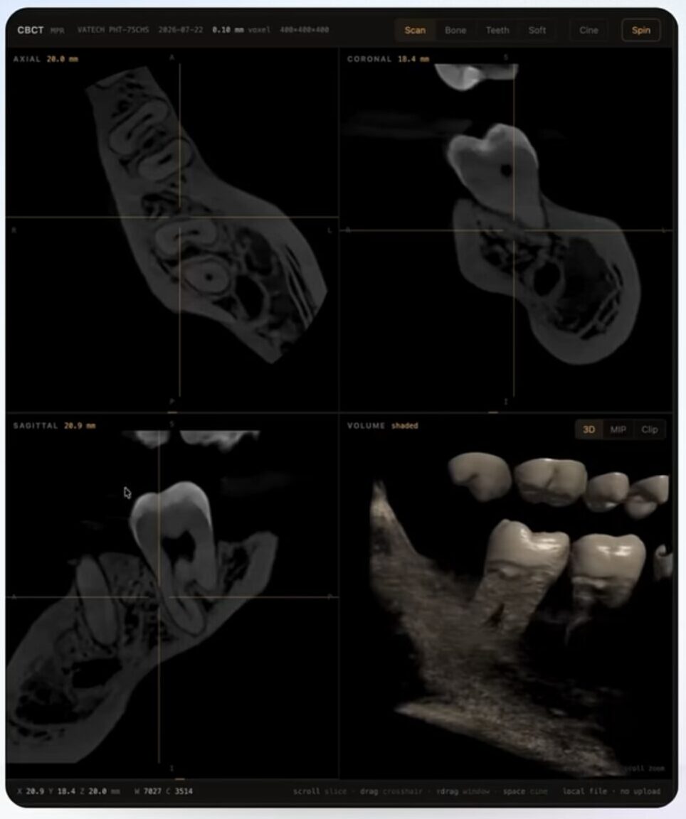
Based on the case report published by the devcode.ai community, instead of spending money for commercial software licenses, Paolo utilizes AI coding assistant Claude Code to build interactive radiology viewers from scratch. This experiment proves how generative artificial intelligence is now able to bridge the technical barriers between clinical data format and ease of access for lay users.
Unravel 800 DICOM files via natural language commands
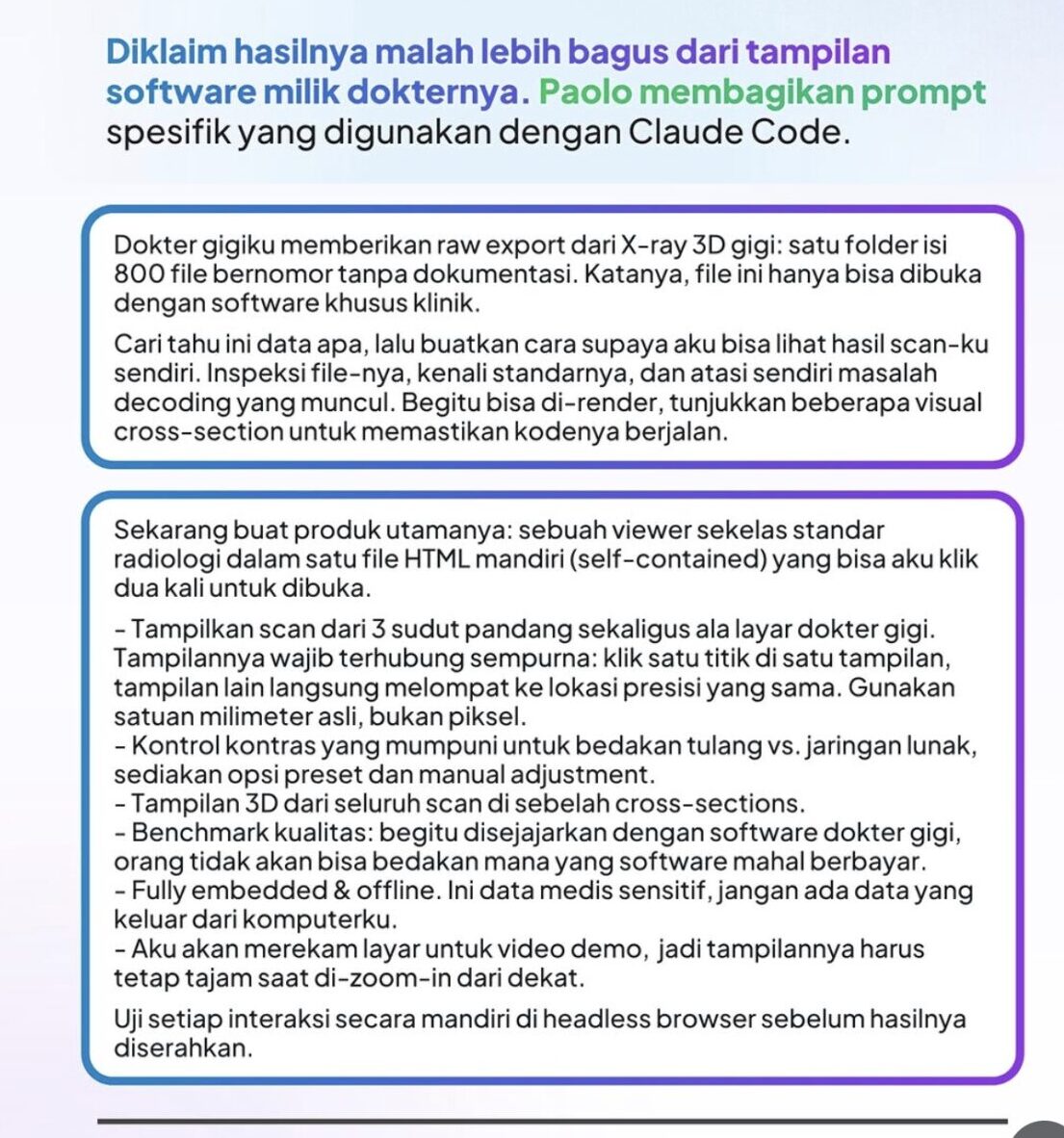
The main challenge in reading CBCT (Cone Beam Computed Tomography) data lies in its file structure which amounts to hundreds of slices without interface documentation. Paolo divides the development process into two stages of structured instruction engineering:
Phase 1: Investigation and Decoding Data Format
In the early stages, Paolo instructed Claude Code to act as a data engineer: inspecting the folder structure, reading the binary metadata DICOM, and solving the image decoding constraint. Claude Code successfully identifies the spatial inter-file coordinates and renders the initial visual chunks (Cross-Section) to verify that the data matrix is read correctly.
Phase 2: Single-file radiology standard viewer creation
Once the decoding logic is proven to be valid, the instructions are directed to construct a complete clinical web interface into the Single self-contained HTML file. This application is designed so that it can be opened with just a double click in a modern web browser without the need for complicated dependency installations or external backend servers.
Comparison: Viewer made by AI vs Commercial Radiology Software
The final result of the viewer made by Paolo is considered capable of presenting very competitive functionality when compared to paid visualization software.


| Parameter | paid clinical radiology software | Custom Viewer via Claude Code |
|---|---|---|
| License fee | expensive (hundreds to thousands of dollars/licensing) | Free (local/independent AI token fee) |
| Distribution Model | Installation of native software (heavy, need admin permission) | Single File Interactive (Portable & Lightweight) |
| Multi-angle navigation | Axial, coronal, sagittal cross-section synchronized | 3 millimeter precision synchronized viewing angle |
| 3D visualization | 3D Volume Shading & Network Segmentation | Integrated 3D Volume Render Interactive |
| Security & Privacy | Depends on the vendor’s cloud policy | 100% offline & private (no data out) |
Practical Steps to Develop a Medical Image Reconstruction Prompt
For developers and users who want to replicate this approach, here is the flow of instructional engineering methodologies that are proven to be effective:
- File deconstruction instructions: Command AI identify raw format and solve binary parsing error before designing the interface.
- Define standard view: Request 3 view of the synchronous panel (axial, coronal, sagittal) where the cursor shift at one corner automatically updates the other corner position.
- Apply windowing/contrast control: Include a contrast adjustment feature (Hounsfield Units/Leveling) to distinguish the structure of hard bones and soft tissue manually or preset.
- Data Privacy Key: Include strict limits that all WebGL/Canvas processing must run locally in the browser memory without any external network connection.
 Medical Record Privacy: Why Offline Architecture Is So Crucial
Biological scanning data such as dental X-rays and skulls contain sensitive biometrics. The Paolo approach that requires the HTML code to run Fully embedded and offline Ensure that medical record data is never sent to third-party servers or used to train public AI models. This experiment is becoming the new standard of how AI agent technology can be utilized to empower ownership of personal data safely and transparently.